| Major Groups > Toothed Mushrooms > Radulomyces copelandii |

|
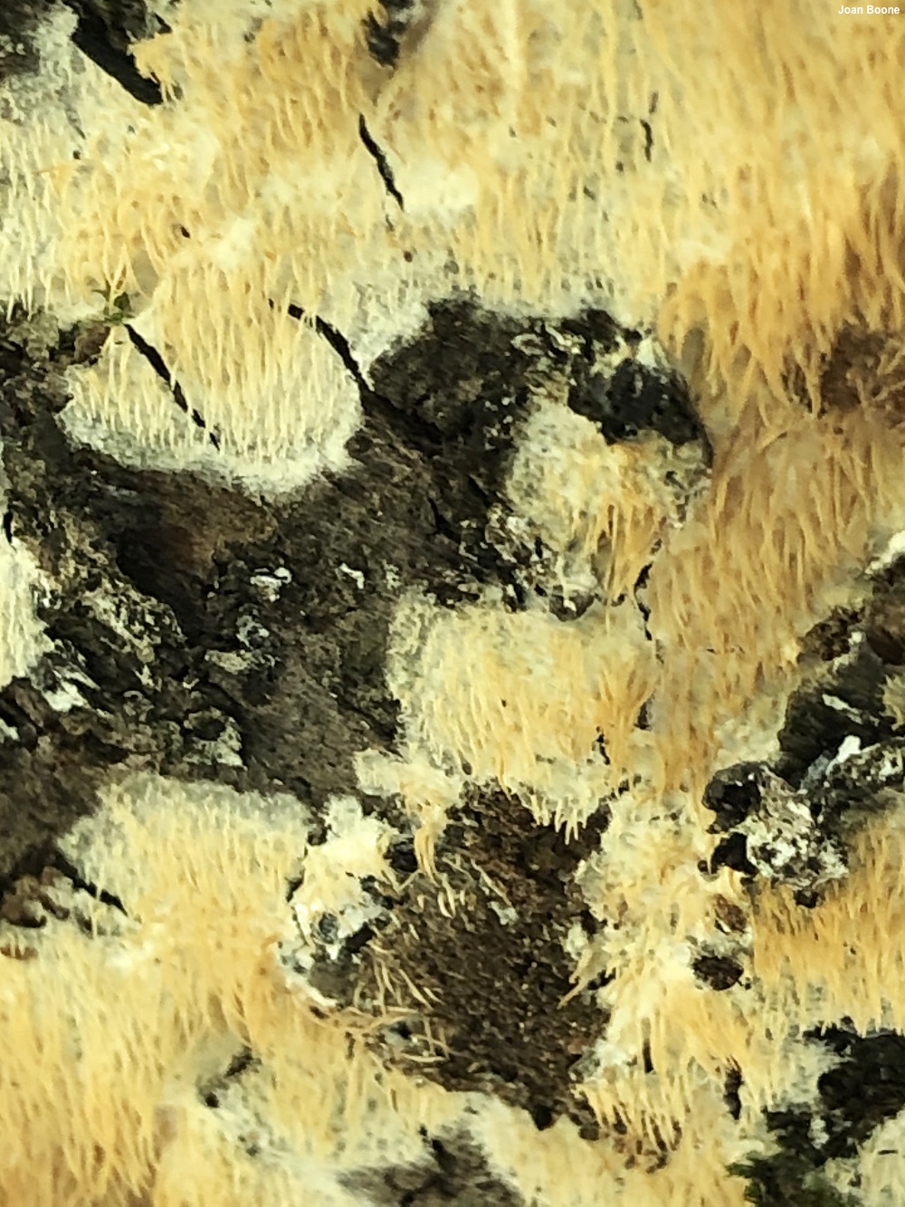
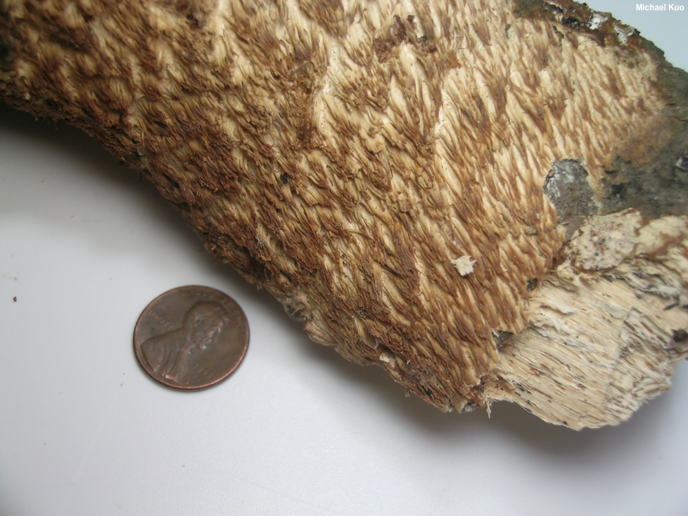
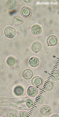
Radulomyces copelandii [ Basidiomycota > Agaricales > Radulomycetaceae > Radulomyces . . . ] by Michael Kuo Found in Asia and in the Mid-Atlantic and northeastern United States, this interesting fungus spreads in resupinate patches on the wood of hardwoods, developing small, clustered spines that are about a centimeter long. The fungus is whitish at first, but becomes yellowish and eventually brownish with age. The spines arise from a very thin mat of tissue—a "subiculum," in Mycologese—and the presence of this layer (which can be hard to see when the fungus is young) can be an important identifier, since some species of Mucronella can look virtually identical but consist of spines that arise directly from the wood, rather than from a subiculum. Radulomyces paumanokensis, described from Long Island, turns out to be a synonym for Radulomyces copelandii (Nakasone et al. 2021); it was described as a separate species since it featured thick clumps of tightly-packed spines that are often flattened and/or branched. Radulodon copelandii is a former name. Thanks to Joan Boone for collecting, documenting, and preserving Radulomyces copelandii for study; her collection is deposited in The Herbarium of Michael Kuo. Description: Ecology: Saprobic; growing on the standing or fallen deadwood of hardwoods, generally when bark is still attached, in cracks and fissures; causing a white rot; summer through winter; originally described from the Philippines; distributed widely from tropical Asia northward to Japan and Russia; in North America recorded in the northeastern and Mid-Atlantic United States. The illustrated and described collection is from Virginia. Fruiting Body: A mass of independent hanging spines arising from a thin layer of tissue (a "subiculum") that spreads across the wood in resupinate patches. Subiculum: Very thin (less than 1 mm thick); pale whitish, becoming yellowish and eventually orangish yellow to brownish with age. Spines: 4–10 mm long; 0.5–1 mm thick at the base; unbranched; bald; at first whitish and proportionally thick-fleshed, becoming yellowish to orangish yellow and narrower, with sharper apices, with age. Dried Specimens: Yellowish to brownish overall. Odor: Not distinctive. Microscopic Features: Spores 5–7 µm; globose to subglobose, with a small apiculus; smooth; hyaline and often uniguttulate in KOH; inamyloid. Basidia 28–34 x 5–8 µm; clavate; 4-sterigmate. Cystidia not found. Hyphal system monomitic; spine tramal hyphae 2–3 µm wide, smooth, thin-walled, hyaline in KOH, with clamp connections. REFERENCES: (N. T. Patouillard, 1914) K. Hjortstam & B. M. Spooner, 1990. (Hjortstam et al., 1990; Nakasone, 2001; Gilbertson & Nakasone, 2003; Ginns & Millman, 2011; Zmitrovich et al., 2017; Wang et al., 2018; Leal-Dutra et al., 2020; Nakasone et al., 2021.) Herb. Kuo 10182002. This site contains no information about the edibility or toxicity of mushrooms. |
© MushroomExpert.Com |

|
Cite this page as: Kuo, M. (2022, March). Radulomyces copelandii. Retrieved from the MushroomExpert.Com Web site: http://www.mushroomexpert.com/radulomyces_copelandii.html |