| Major Groups > Gilled Mushrooms > Phylloporus & Phylloporopsis |
|
|
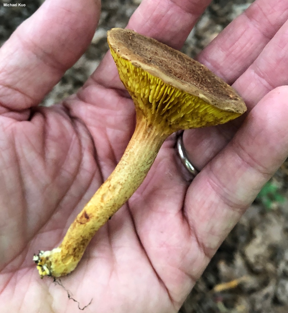
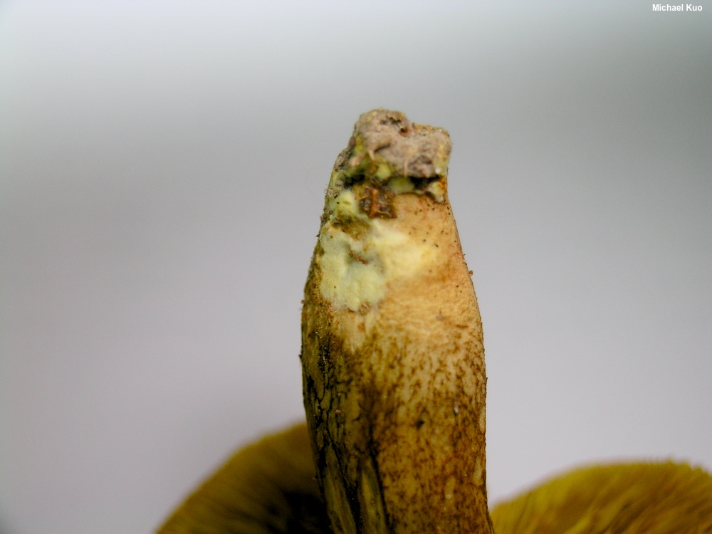
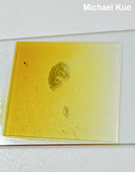
[ Basidiomycota > Boletales > Boletaceae . . . ] Phylloporus and Phylloporopsis by Michael Kuo, 24 October 2025 This is a small group of rather odd mushrooms, often called the "gilled boletes." From above, they look for all the world like boletes. But turn them over and you'll find gills where you expected pores. The stems are bolete-ish, the chemical reactions are similar, and some gilled boletes even stain blue, like some boletes. The similarity continues under the microscope; gilled bolete spores are more or less spindle-shaped and look rather like the spores of most boletes, while cystidia and pileipellis elements are also bolete-like. Currently, identifying the various gilled boletes in the United States and Canada is fairly straightforward, and can be accomplished with observation of physical features; microscopic analysis is probably not needed, especially if you have collected fresh specimens in good condition. But the sample size for DNA-supported studies is very low, and "new" phylogenetic species may be hanging out across the continent, in the woods and in herbaria. In Mexico several species have recently been separated; see the references list below for the relevant papers. There are a couple of pitfalls to bear in mind as you attempt to identify gilled boletes: blue staining, and the color of the basal mycelium. Blue staining of the gills and flesh in the gilled boletes is not as stable as some field guides (and even some technical treatments) would have you believe. Most of the species are capable of blue staining—though some, perhaps, do so more frequently. Thus Phylloporus rhodoxanthus does not usually stain blue, but it does occasionally, while the opposite is the case for Phylloporus leucomycelinus, which usually stains blue but occasionally does not. The color of the basal mycelium, either yellow or white, is a useful character for separating some of the gilled boletes, but yellow basal mycelium can fade to whitish in older, dried-out specimens. More confusing, however, is the white fuzz created by a Hypomyces that frequently attacks gilled boletes, beginning at the base of the stem. This is a fairly commmon illusion, to judge from my collections, and while the Hypomyces (it is probably Hypomyces microspermus) is sometimes clearly a Hypomyces, it can also look for all the world like basal mycelium, especially when it is just beginning to develop. I have provided some illustrations of this illusion to the left. Fortunately for those who have access to microscopes, the true state of affairs can be sorted out quickly by examining a bit of the putative mycelium; if it is actually a Hypomyces, corresponding hyphal structures and aleuriospores will be present. Some sources refer to the "fleeting amyloid reaction" that can be observed in some of the gilled boletes. Although assessing this reaction does not require a microscope, it does require access to Melzer's reagent (whether or not the much more accessible Lugol's solution would work as a substitute has not been determined, to my knowledge). Mount a fresh or rehydrated piece of a gill in a drop or two of Melzer's on a slide, and press down a coverslip with a pencil eraser. In many gilled boletes the gill tissue will soon develop an amyloid, dark blue to dark bluish gray, reaction. The reaction, which is also known as "Imler's reaction," disappears within a few minutes. I have not found assessing this reaction to be particularly helpful in identifying my North American collections. Species of Phylloporus are so closely related to boletes in the genus Xerocomus (see Xerocomus subtomentosus for an example) that the genera should be merged, in my humble opinion, as some European authors (e.g. Bresinsky & Binder 2003, Noordeloos 2018) have done. Results from multiple contemporary, DNA-based phylogenies support this idea (see Binder & Hibbet 2006, Nuhn et al., 2013, Wu et al. 2014, Kuo & Ortiz-Santana 2020); the way the phylogenetic trees branch in these studies makes Xerocomus untenable if Phylloporus is maintained, and one is forced to create a series of confusing new genera—or to combine them tidily as Xerocomus, along with the recently erected genus Hourangia. The latter option is much better because it simplifies the taxonomy and, most importantly, better communicates the close evolution of the organisms through the shared genus name. Unfortunately most of the North American Phylloporus species have not yet been combined as species of Xerocomus, so I am forced to treat them in Phylloporus—and while Xerocomus rhodoxanthus is a legitimate coimbination, I will suck it in and treat it as Phylloporus rhodoxanthus to avoid confusion. Key to 6 American and Canadian Phylloporus and Phylloporopsis Species
Phylloporus foliiporus . . . was named by Murrill (1943, as Gomphidius foliiporus) on the basis of a collection made "under an oak" in the Gainesville, Florida area. Apparently Murrill's collection, along with a second collection made the next day, had a very dark spore print; he described the color as "pale-umbrinous, umbrinous, or purplish-brown." Murrill was confused by the mushroom, and wrote that it was "one of those puzzles that causes loss of sleep. When first sighted I thought of Stropharia, but the gills were too decurrent. Although showing kinship with Phylloporus and Paxillus, the spores were far too dark. I am placing it with Gomphidius temporarily because there is no other place for it to go, brushing aside for the moment several obvious and good reasons for not doing so." The species has been variously interpreted over the years, usually as a bluing species with yellow basal mycelium. But Phylloporus rhodoxanthus occasionally features blue staining, and such specimens may well account for much of what has been called Phylloporus foliiporus. According to Neves & Halling (2010), "[o]ne of the most diagnostic characteristics of this taxon is the presence of hymenial cystidia with a melleous-colored apex, sometimes constricted." The authors examined a total of seven collections, from Florida, Alabama, and Japan, and apparently found the honey colored tips on all the cystidia they observed. They did not specify the mounting medium, unfortunately, though it was probably KOH. Among the seven collection examined by Neves and Halling was one made by J. L. Mata in Alabama in 2005 (Mata 1677, held in New York); this collection appears in a 3-gene phylogeny created by Gutiérrez-Domínguez et al. (2024), alongside another foliiporus collection made by S. Jakob (iNAT 86166978) in Pompano Beach. Photos of these two collections can be found online with a little digging. Unfortunately Murrill's type collection was not also sequenced by Gutiérrez-Domínguez, so we do not know whether the hypothesis of Neves & Halling—that Mata's collection and the type collection of foliiporus are the same—is supported or not. References Farid, A. & 11 coauthors (2018). Phylloporus and Phylloboletellus are no longer alone: Phylloporopsis gen. nov. (Boletaceae), a new smooth-spored lamellate genus to accommodate the American species Phylloporus boletinoides. Fungal Systematics and Evolution 2: 341–359. Gutiérrez-Domínguez, E., L. Montoya, A. Ramos, A. Andrade-Torres, J. C. Noa-Carrazana, Á. I. Ortiz-Ceballos & V. M. Bandala (2024). Two new species of Phylloporus (Boletaceae) from the montane cloud forest of eastern Mexico. Phytotaxa 668: 44–62. Kuo, M. & B. Ortiz-Santana (2020). Revision of leccinoid fungi, with emphasis on North American taxa, based on molecular and morphological data. Mycologia 112: 197–211. López, A. R. & J. García (2018). Phylloporus foliiporus. Funga Veracruzana 166. Montoya, L. & V. M. Bandala (1991). Studies on the genus Phylloporus in Mexico, I. Discussion of the known species and description of a new species and a new record. Mycotaxon 41: 471–482. Montoya, L. & V. M. Bandala (2011). A new Phylloporus from two relict Fagus grandifolia var. mexicana populations in a montane cloud forest. Mycotaxon 117: 9–18. Montoya, L., E. Garay-Serrano & V. M. Bandala (2019). Two new species of Phylloporus (Fungi, Boletales) from tropical Quercus forests in eastern Mexico. MycoKeys 51: 107–123. Neves, M. A. & R. E. Halling (2010). Study on species of Phylloporus I: Neotropics and North America. Mycologia 102: 923–943. Neves, M. A., M. Binder, R. Halling, D. Hibbett & K. Soytong (2012). The phylogeny of selected Phylloporus species, inferred from NUC-LSU and ITS sequences, and descriptions of new species from the Old World. Fungal Diversity 55: 109–123. Singer, R., C. L. Ovrebo & R. E. Halling (1990). New species of Phylloporus and Tricholomopsis from Colombia, with notes on Phylloporus boletinoides. Mycologia 82: 452–459. Tremble, K. & 9 coauthors (2024). A revised phylogeny of Boletaceae using whole genome sequences. Mycologia 116: 392–408. Wu, G., B. Feng, J. Xu, X.-T. Zhu, Y.-C. Li, N.-K. Zeng, Md. I. Hosen & Z. L. Yang (2014), Molecular phylogenetic analyses redefine seven major clades and reveal 22 new generic clades in the fungal family Boletaceae. Fungal Diversity 69: 93–115. This site contains no information about the edibility or toxicity of mushrooms. Kuo, Michael (2025, October). Phylloporus and Phylloporopsis. Retrieved from the Mushroomexpert.Com website: www.mushroomexpert.com/phylloporus.html All text and images © , Mushroomexpert.com |