| Major Groups > Mycotrophs > Pseudoboletus parasiticus |
| Major Groups > Boletes > Pseudoboletus parasiticus |

|
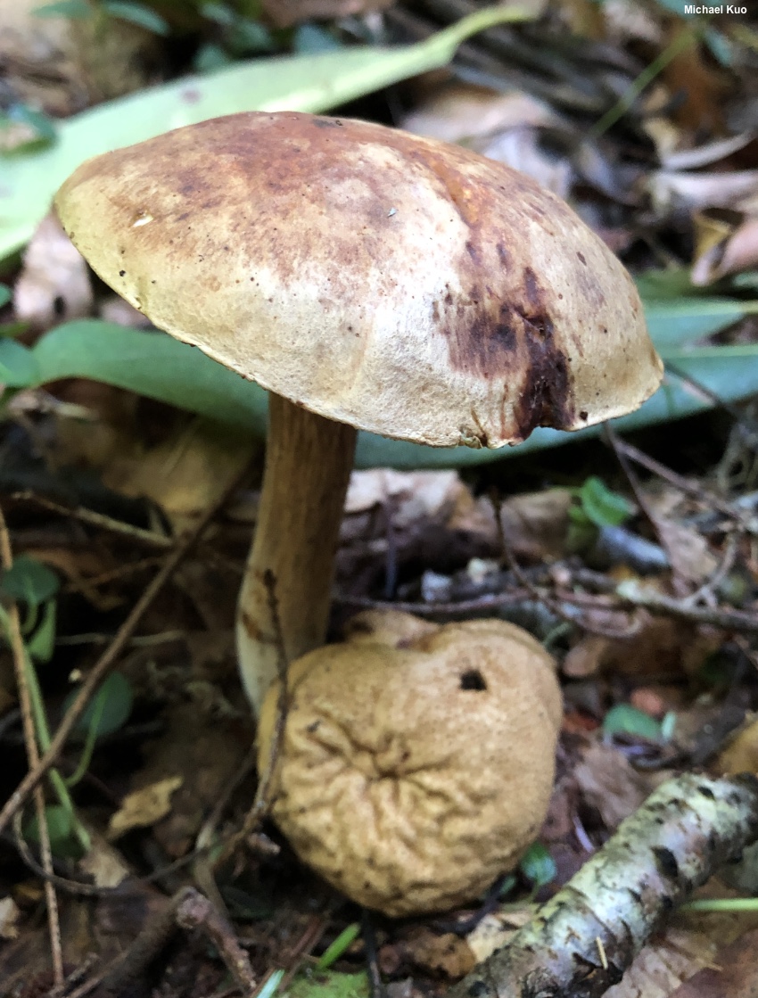
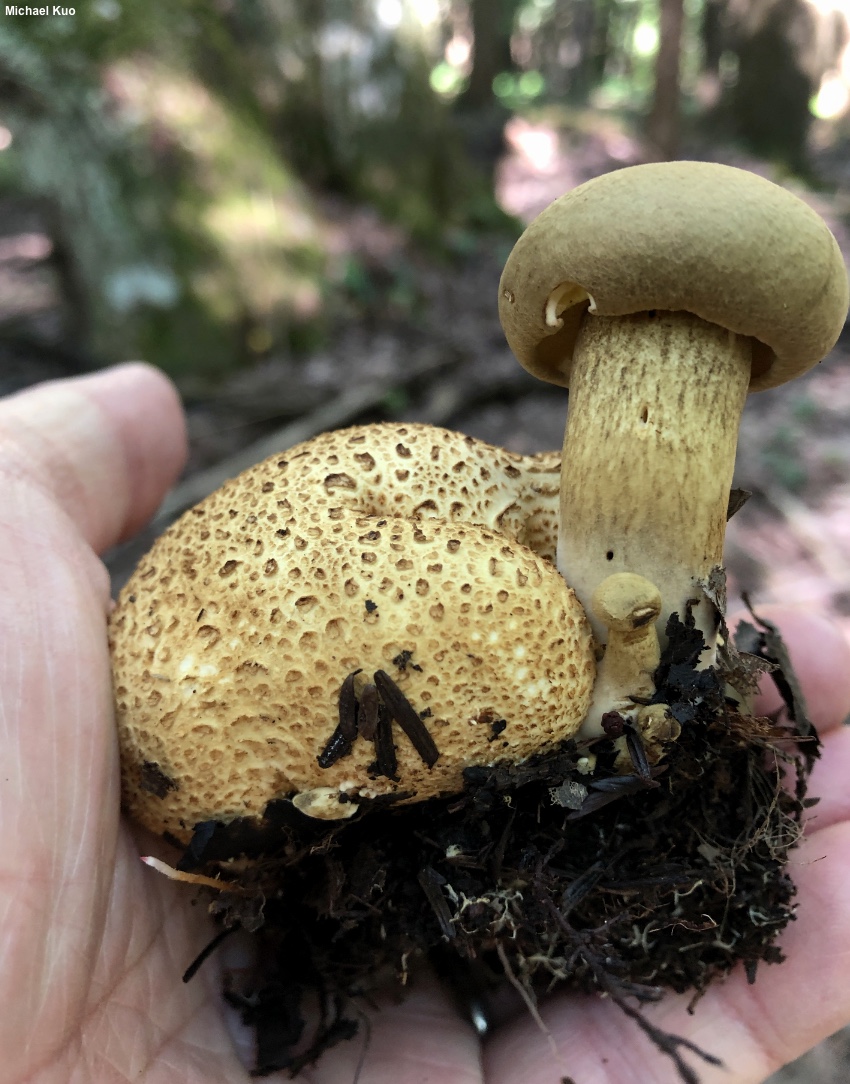
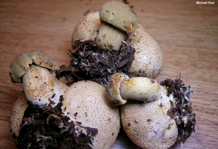
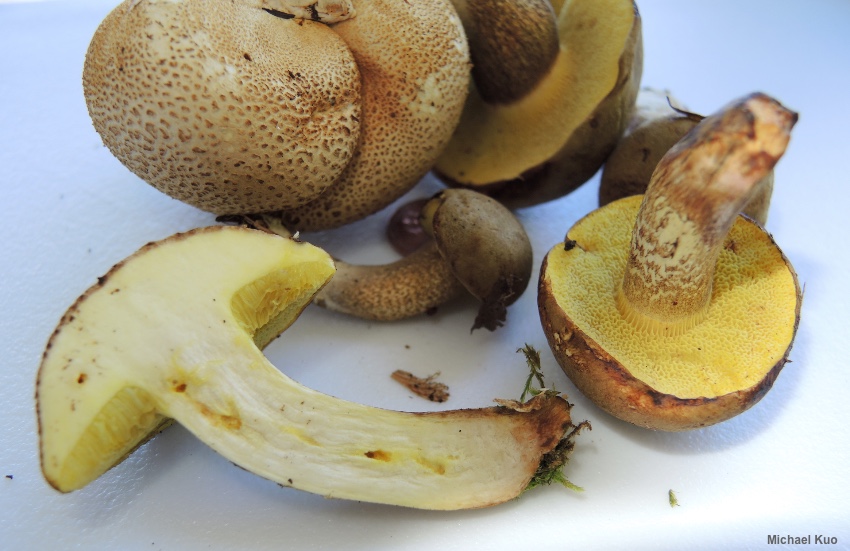
Pseudoboletus parasiticus [ Basidiomycota > Boletales > Boletaceae > Pseudoboletus . . . ] by Michael Kuo Here is one bolete species, at least, that is a cinch to identify: it parasitizes the earthball Scleroderma citrinum, growing right out of its body! There are many other examples of mushrooms parasitizing other mushrooms (see the key to mycotrophs for examples), but none of the other cases involves a bolete popping out of an earthball. If you were to separate Boletus parasiticus from its host, however, you would have a pretty nondescript little bolete with a yellowish brown cap and a honey-yellow, xerocomoid pore surface. In this case the red reaction of the cap to the application of a drop of ammonia might help distinguish the mushroom, as would the presence of many tiny, dark fibers on the stem, which almost look like miniature versions of the scabers found in Leccinum species. Under the microscope Pseudoboletus parasiticus has spores, cystidia, and pileipellis elements similar to those of many other species—but the stipitipellis (the surface of the stem) differs from that of other boletes in its lack of caulohymenium; no basidia and cystidia can be found in patches on the stem, even directly below the pore surface. This difference led Šutara (1991) to place the species in its own genus, Pseudoboletus, and the idea has been supported more recently by DNA studies (e.g. Nuhn et al. 2013, Wu et al. 2016). How does this mushroom survive? What I mean is, after it has distributed its spores, what happens to them? Do they float around in the air currents, waiting for earthballs to appear so they can dive-bomb and germinate? Do they land in substrate that contains earthball mycelium, then camp out, waiting for the earthball organism to make fruiting bodies? Or does Pseudoboletus parasiticus parasitize Scleroderma citrinum even in the mycelium—do the mycelia of the two species grow together? I first typed these questions in 2003, in an earlier version of this webpage. Since then definitive answers have not surfaced, to my knowledge, although one recent source (Læssøe & Petersen 2019) speculates as follows:
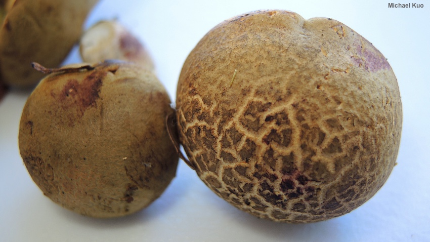
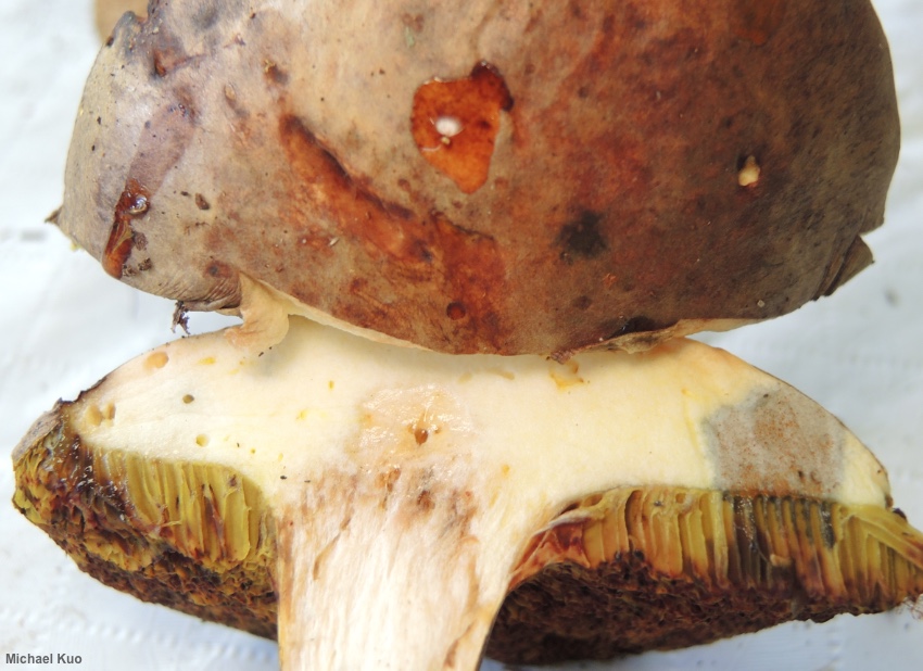
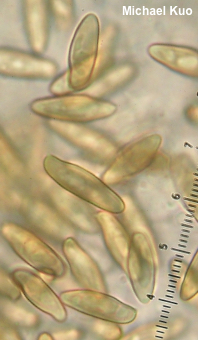
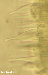
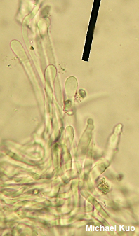
However, the situation is further complicated by the fact that researchers (Richter & Bruhn 1989) have been able to grow mycorrhizae in culture between Pseudoboletus parasiticus and Pinus resinosa (red pine)—raising the possibility that Pseudoboletus parasiticus is actually mycorrhizal. Xerocomus parasiticus is a synonym, as is Boletus parasiticus. Description: Ecology: Apparently parasitic on Scleroderma citrinum, which is mycorrhizal with hardwoods and conifers (but see the discussion above); most frequently found, in my collecting experience, when the host is growing in eastern hemlock bogs; growing alone or in small clusters; summer and fall; originally described from France, and widespread in Europe; in North America distributed fairly widely east of the Great Plains. The illustrated and described collections are from Kentucky, Michigan, and North Carolina. Cap: 2.5–7 cm across; convex, becoming broadly convex; dry; suedelike; becoming cracked with age; yellowish brown with a hint of olive; sometimes bruising reddish; margin rolled under when young. Pore Surface: Yellow, becoming olive yellow and, eventually, brown to reddish brown; not bruising; xerocomoid; with 1–2 angular pores per mm; tubes 3–6 mm deep. Stem: 4–8 cm long; 1–2 cm thick; equal; often curved near the base; dry; solid; colored more or less like the cap; covered with fine brown aggregations of fibrils, often in chevron patterns; sometimes bruising reddish when handled; basal mycelium white. Flesh: Pale yellow, turning slowly chrome yellow above the tubes and in the stem; reddish in the stem base. Odor and Taste: Not distinctive. Chemical Reactions: Ammonia red to pink on cap surface; negative on flesh. KOH orangish on cap surface; negative to orangish on flesh. Iron salts negative on cap surface; blue-gray on flesh. Microscopic Features: Spores 13–21 x 4–6 µm; boletoid-fusiform; smooth; golden in KOH. Basidia 35–40 x 5–7.5 µm; subclavate; 4-sterigmate. Hymenial cystidia 50–75 x 7.5–15 µm; fusiform or lageniform, with a long neck; smooth; thin-walled; hyaline in KOH. Pileipellis a collapsing trichoderm; elements 5–7.5 µm; wide, smooth, hyaline to yellowish in KOH; terminal cells cylindric with rounded to subclavate apices. Stipitipellis a collapsing trichoderm; elements 3–8 µm wide, smooth, hyaline to yellowish in KOH; terminal cells ranging from cylindric with rounded apices to subclavate or fusiform. Caulohymenium not found. REFERENCES: (Bulliard, 1790) Šutara, 1991. (Fries, 1821; Saccardo, 1888; Coker & Beers, 1943; Singer, 1945; Snell & Dick, 1970; Smith & Thiers, 1971; Grund & Harrison, 1976; Phillips, 1981; Smith, Smith & Weber, 1981; Weber & Smith, 1985; Richter & Bruhn, 1989; Breitenbach & Kränzlin, 1991; Phillips, 1991/2005; Lincoff, 1992; Both, 1993; Barron, 1999; Bessette, Roody & Bessette, 2000; Roody, 2003; McNeil, 2006; Miller & Miller, 2006; Kuo, 2007; Nonis, 2007; Binion et al., 2008; Šutara, 2008; Buczacki et al., 2012; Nuhn et al., 2013; Kuo & Methven, 2014; Bessette et al., 2016; Wu et al., 2016; Baroni, 2017; Gminder & Böhning, 2017; Elliott & Stephenson, 2018; Knudsen & Taylor, 2018; Noordeloos, 2018; Sturgeon, 2018; Læssøe & Petersen, 2019.) Herb. Kuo 09029503, 08180606, 08091917, 08112005. This site contains no information about the edibility or toxicity of mushrooms. |
© MushroomExpert.Com |


|
Cite this page as: Kuo, M. (2020, October). Pseudoboletus parasiticus. Retrieved from the MushroomExpert.Com Web site: http://www.mushroomexpert.com/pseudoboletus_parasiticus.html |