| Major Groups > Gilled Mushrooms > Phylloporus & Phylloporus > Phylloporus rhodoxanthus |

|
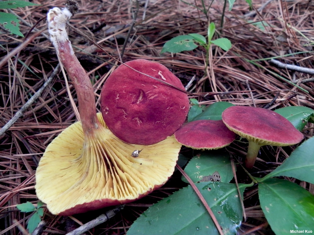
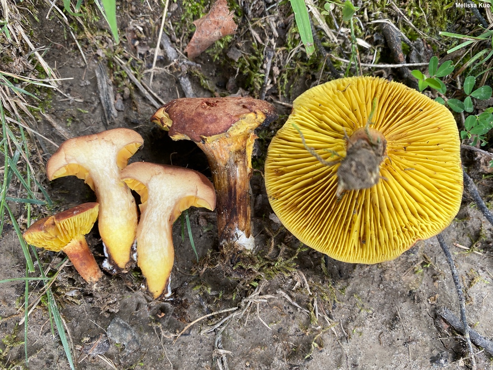
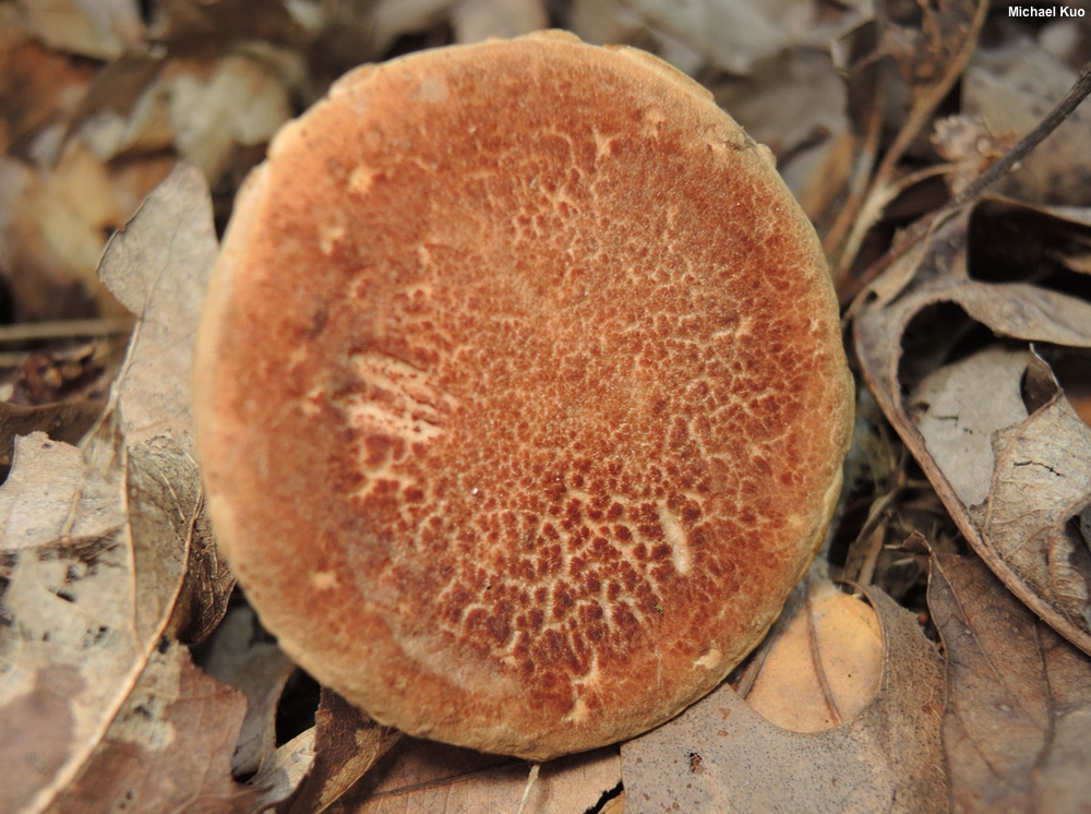
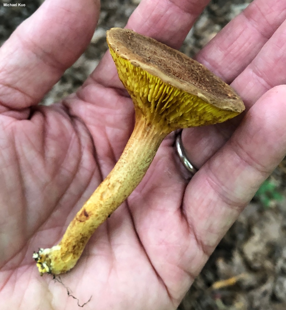
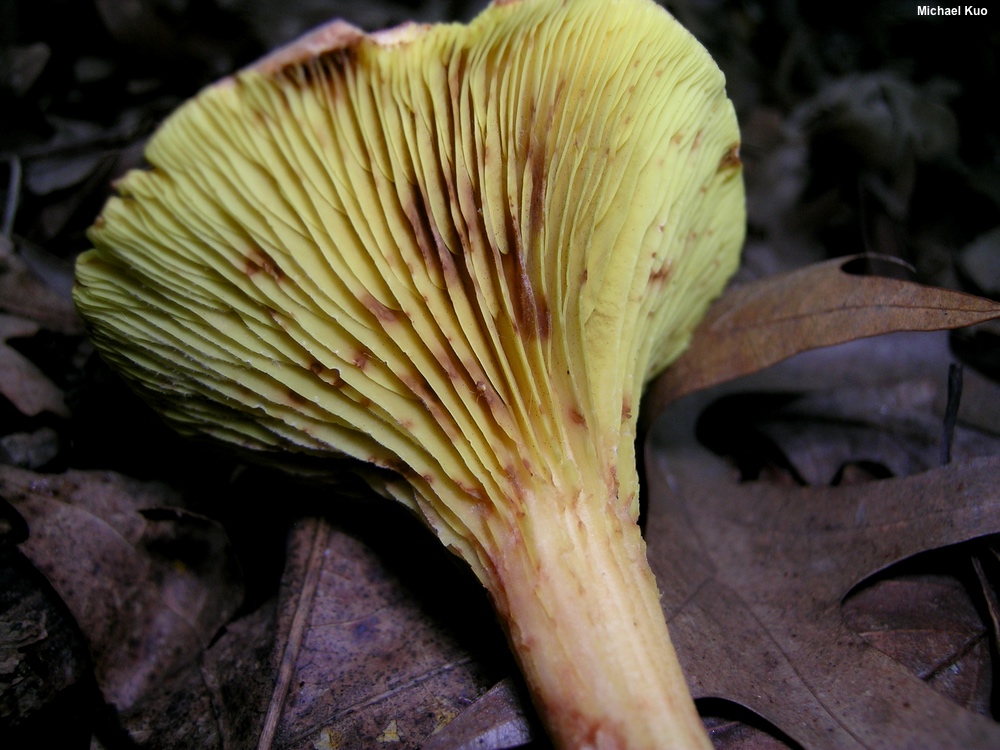
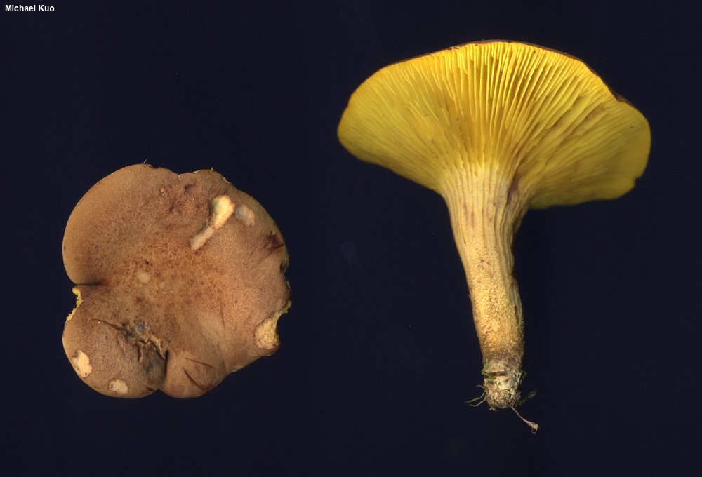
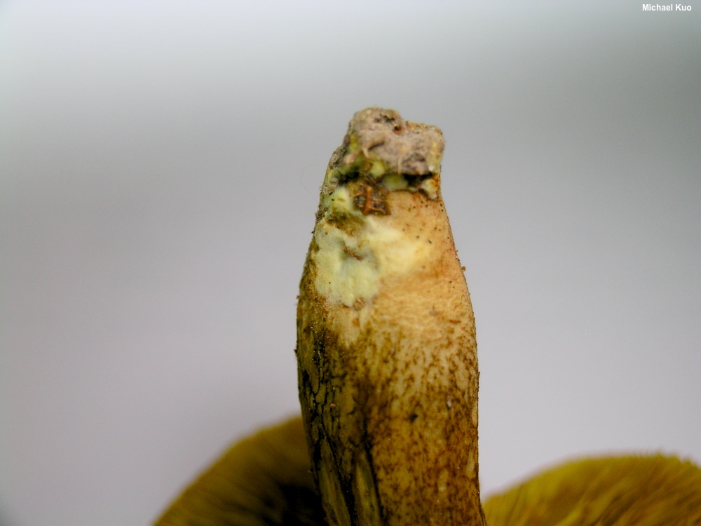
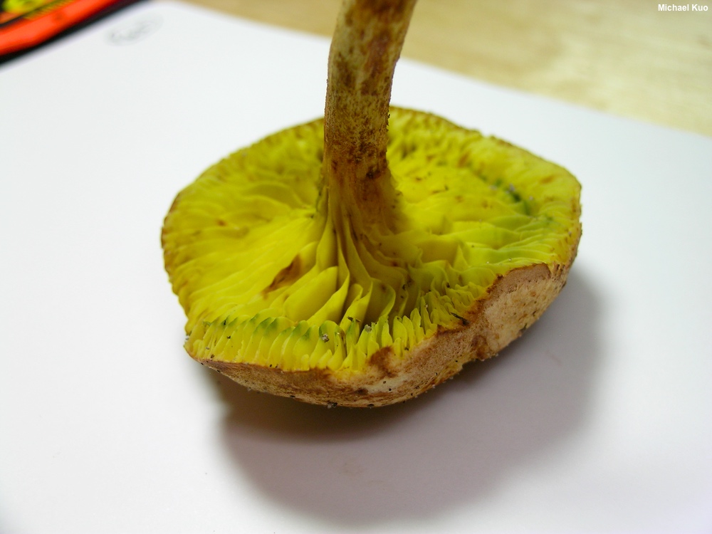
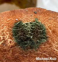
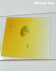
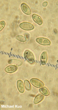
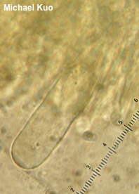
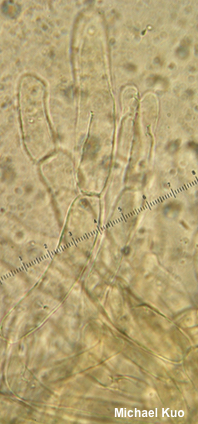
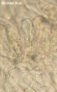
Phylloporus rhodoxanthus [ Basidiomycota > Boletales > Boletaceae > Phylloporus . . . ] by Michael Kuo, 23 October 2025 This common summer gilled bolete appears in hardwood forests east of the Rocky Mountains. Its colors range from red to brown (usually somewhere between), and its gills, when fresh, are bright yellow. The base of the stem features yellow basal mycelium, and the cap turns green with a drop of ammonia. The ammonia reaction is probably not needed for successful identification, but it's fun. Compare Phylloporus rhodoxanthus with Phylloporus leucomycelinus, which is virtually identical but features white basal mycelium, and with Phylloporopsis boletinoides, which has dull yellowish gills that mature to brownish, and is associated with hard pines in the southeastern United States. Also compare with Phylloporus 01, which is orange and differs in its pileipellis. The yellow basal mycelium of Phylloporus rhodoxanthus is often obscured by a white Hypomyces (probably Hypomyces microspermus) that starts to develop at the stem base, leading to confusion with Phylloporus leucomycelinus. See the examples among the photos to the right. According to most sources Phylloporus rhodoxanthus does not stain blue when sliced open or when the gills are bruised, but in my collecting experience this is not completely accurate: occasionally this species does stain blue, though it is not the norm. The best name for this fungus is Xerocomus rhodoxanthus, but I am treating it as a Phylloporus to avoid confusion. See the discussion on the page for gilled boletes if you care. Thanks to Django Grootmyers for documenting, collecting, and preserving Phylloporus rhodoxanthus for study; his collection is deposited in The Herbarium of Michael Kuo. Description: Ecology: Mycorrhizal with oaks and other hardwoods; growing alone or gregariously; summer; originally described from North Carolina (von Schweinitz 1822); widely distributed in North America east of the Rocky Mountains. The illustrated and described collections are from Illinois, Indiana, and Ohio. Cap: 2–8.5 cm (occasionally even larger, to 13 cm); convex, becoming broadly convex, flat, or shallowly depressed; dry; bald or finely velvety; sometimes developing a fine network of cracks; reddish to reddish brown, brown, or yellowish brown. Gills: Running down the stem; nearly distant; thick; short-gills frequent; bright yellow, becoming rusty-spotted; not usually bruising but occasionally bruising blue. Stem: 3–8 cm long; up to 1 cm thick; tapered downward; often with extending "gill lines" at the apex; yellowish, with reddish to brown fibrils; basal mycelium yellow. Flesh: White to pale yellow; not usually staining when exposed but sometimes bluing faintly when sliced. Odor and Taste: Not distinctive. Chemical Reactions: Ammonia blue-green on fresh cap surface (sometimes flashing green and resolving to red, purple, or blackish); red on dried cap surface. KOH black, negative, or orangish on cap; pinkish orange on flesh. Iron salts negative to gray on cap; negative to pale bluish green on flesh. Fleeting amyloid reaction positive for gill tissue. Spore Print: Olive brown. Microscopic Features: Spores 8–11 x 3.5–5 µm; subfusiform or occasionally subellipsoid; smooth; hyaline to yellowish in KOH. Basidia 32–38 x 5–7 µm; clavate; 4-sterigmate. Pleurocystidia 40–90 x 10–16 µm; widely cylindric to subfusiform, utriform, or lageniform (rarely sphaeropedunculate); smooth, or sometimes apically encrusted (best seen with Melzer's reagent); thin- or thick-walled; hyaline in KOH. Pileipellis a trichoderm; elements cylindric, 6–14 µm wide, smooth or finely roughened; hyaline to yellowish in KOH; terminal cells cylindric with rounded or subclavate apices; subterminal cells cylindric. Stipitipellis a layer of repent, parallel, cylindric hyphae 3–7 µm wide, smooth, walls up to 1.0 µm thick, yellowish in KOH; caulohymenium in tufts, caulocystidia similar to pleurocystidia, often clavate. Clamp connections not found. REFERENCES: (L. D. von Schweinitz, 1822) G. Bresàdola, 1900. (Kauffman, 1918; Smith, 1975; Smith, Smith & Weber, 1979; Weber & Smith, 1985; Arora, 1986; Phillips, 1991/2005; Lincoff, 1992; Horn, Kay & Abel, 1993; Barron, 1999; Roody, 2003; Miller & Miller, 2006; Kuo, 2007; Binion et al., 2008; Neves & Halling, 2010; Kuo & Methven, 2014; Baroni, 2017; Woehrel & Light, 2017; Elliott & Stephenson, 2018; Farid et al., 2018; McKnight et al., 2012; Gutiérrez-Domínguez et al., 2024; Tremble et al., 2024.) Herb. Kuo 07089503, 08300215, 07160403, 07130805, 06211003, 07221003, 06301102, 07131402, 07101604, 07141609, 07191601, 06221804, 08052004, 07172103, 07302401. This site contains no information about the edibility or toxicity of mushrooms. |
© MushroomExpert.Com |
|
Cite this page as: Kuo, M. (2025, October). Phylloporus rhodoxanthus. Retrieved from the MushroomExpert.Com Web site: http://www.mushroomexpert.com/phylloporus_rhodoxanthus.html |